Mohs Surgery

Mohs micrographic skin cancer surgery or simply “Mohs surgery” is named after the surgeon, Dr. Frederic E. Mohs, who developed this innovative procedure. This precise form of skin cancer excision is the gold standard for removal of some of the most challenging skin malignancies located in the most sensitive regions such as the face, hands, feet and genitals. For these tumors Mohs surgery attains the highest cure rates achievable with studies demonstrating skin cancers are completely eradicated in 97 to 99% of cases.
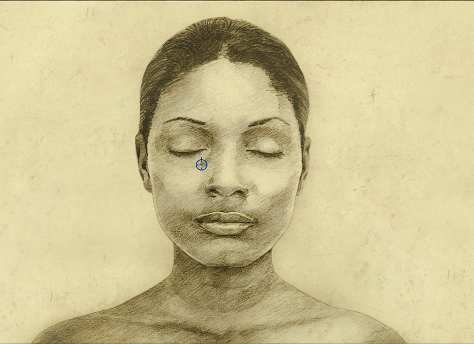
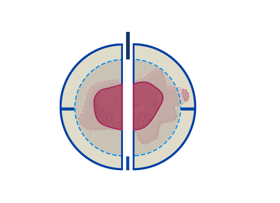
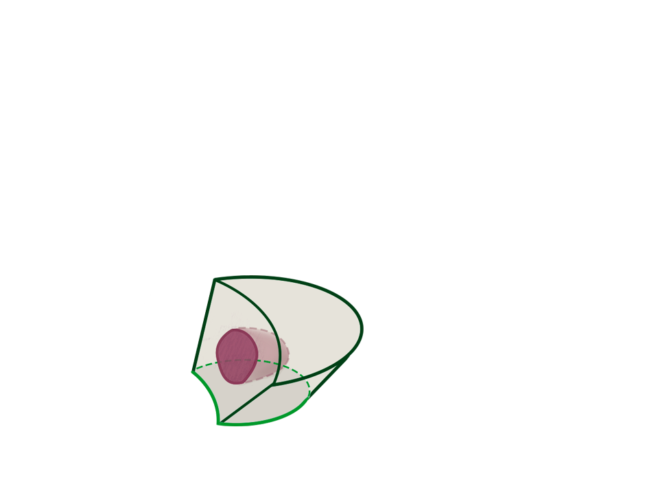
Figure 1
In the illustration to the left, the patient demonstrates a skin cancer in the cosmetically sensitive area below her right eye.


Targeting Skin Cancer

Skin cancers amenable to Mohs surgery include both basal cell and squamous cell carcinoma, though some forms of melanoma and other less common skin tumors have also been treated with this technique. In preparation for surgery the edges of the visible tumor are demarcated with ink and the area is numbed with local anesthesia.

Figure 2
The patient’s tumor is marked for removal.


Precise Tumor Extraction

The tumor’s orientation within the surrounding skin is tracked with specific markings. This allows a “map” of the tumor to be established that can always be related back to its original position within the patient’s skin.

Figure 3
The first stage of the tumor is removed with care to maintain markings representing its anatomical orientation. The tissue is commonly divided in half for ease of handling and to assist in marking its orientation.


Microscopic Analysis

The success of Mohs surgery is founded in the utilization of the microscope to analyse 100% of the margin of any tissue that is removed. This does away with the need to remove extra healthy tissue, and thus Mohs surgery is highly regarded as a “tissue sparing” technique. Removed tissue is prepared within about an hour to be analysed under a microscope. Once all of the edges have been examined, a determination is made as to whether any additional tissue is required to be removed in order to completely eradicate the cancer.

Figure 4
Skin removed in the first stage is observed under the microscope. The tissue has been divided in half and the peripheral edges are laid flat for examination. 100% of the periphery is examined microscopically.


Tissue Sparing

A second stage of tissue is removed only at the precise point where the map indicates residual tumor remains. This insures that only diseased tissue is eliminated and that only minimal healthy tissue is sacrificed. As many stages as necessary are performed to clear all the cancer. On average, however, only 2 or 3 stages are necessary to attain a complete cure from the skin cancer in most cases.

Figure 5
An additional stage of tissue is removed at the “2:30” position where microscopic tissue analysis demonstrated skin cancer had still been present in this patient.


Fine Cosmetic Closure

Meticulous “margin control” leads to the extremely high rates of curing skin cancer when Mohs surgery is performed. With this level of confidence that the cancer has been eliminated, the affected area can now be reconstructed utilizing the latest cosmetic surgical techniques.

Figure 6
The patient incision is closed with fine cosmetic sutures and techniques to minimize any residual evidence of prior surgery.


Highest Standard of Care

Whenever possible, the surgical closure is placed in the border between different anatomical regions of the face to further minimize its visibility. With the surgical closure in this patient placed along the border between the lower eyelid and the cheek, its appearance is muted. In summary, Mohs surgery incorporates microscopic guided tumor excision along with the sparing of healthy tissue to achieve the highest skin cancer cure rates. This, in combination with the latest cosmetic closure techniques, provides the utmost aesthetic outcomes resulting in a procedure that delivers the current standard of excellence in skin cancer care.

Figure 7
This patient’s closure is already well concealed. Its appearance will typically continue to diminish with time.


Credits

Designed by Rachelle Lortie.
rlortie@ecuad.ca

Information provided by Dr. Charles Lortie.
BSc(Med), MD, CCFP, FCFP, FRCPC, FAAD Board Certified Dermatologist (Canada & USA)
drlortie.com

'Small Crosses' background pattern made by Ian Dmitry, modified by Rachelle Lortie (CC BY-SA 3.0 US).
Avaliable here